Understanding The Anatomy of the Wrist
 The wrist is a complex joint connecting the forearm with the hand. It’s unique makeup allows for the critical range of motion we depend on for all the intricate hand movements we enjoy. We use our wrists extensively and in fact they are involved in everything we do with our hands. Therefore our wrists are prone to many types of injury ranging from repetitive use to impact to arthritic and more.
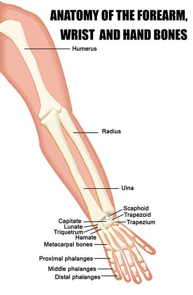
Many bones are involved in the anatomy of the wrist. These include the distal ends of both the ulna and the radius in the forearm and eight carpal bones . Ligaments are an important component of the wrist providing both strength and stability. Each of the many bones of the wrist is connected to another bone by one or more ligaments. Muscles affecting the wrist are located on the forearm with the tendons extending into the wrist and hand. Finally we will take a look at the innervation of the wrist as well to round out our understanding of its anatomy.
 Bones of the Wrist
The distal ends of both the forearm bones, the ulna and the radius articulate with the carpal bones to form the wrist. There are eight carpal bones. They are arranged in two rows, proximal (closer) and distal (further).
Proximal Bones of the Wrist
- Lunate
- Triquetrum
- Pisiform
Distal Bones of the Wrist
- Capitate
- Trapezium
- Trapezoid
- Hamate
Finally, the Scaphoid bone crosses both rows, distal and proximal and is therefore the largest of the carpal bones. It is the scaphoid and the lunate bones which directly articulate with the radius and ulna to create the wrist joint.
A broken wrist is a common injury and may involve one of more of the many bones of the wrist in an actual break or fracture.
Ligaments of the Wrist
There are a large number of ligaments in the wrist. Each of the many bones is attached to another by ligaments. The main and largest of the wrist ligaments are the medial collateral ligament (MCL) and the lateral collateral ligament (LCL). The MCL runs from the distal end of the ulna and crosses the wrist attaching to both the triquetrum bone and the pisiform bone. The LCL passes from the end of the radius bone and crosses the joint to the scaphoid.
Sprains and strains of the wrists are one of the most common injuries. Falls, impact, overuse, arthritis and age may all be factors leading to this common wrist injury including carpal tunnel syndrome.
Muscles Affecting the Wrist
Muscles which act on the wrist are generally located in the forearm with the tendons crossing into the hand. On the back of the forearm are the muscles that extend the wrist or pull it back. These are; extensor carpi radialis brevi, extensor carpi radialis longus, extensor carpi ulnaris, extensor digitorum communis and extensor pollicus longus.
On the front of the forearm are the muscles which allow flexion of the wrist.
These muscles are:
- Flexor carpi radialis
- Flexor carpi ulnaris
- Flexor digitorum superficialis
- Flexor pollicis longus
An injury to any of these muscles will affect the ability of the wrist to perform its normal activity and range of motion.
Nerves of the Wrist
There are three nerves that run from the foreman, across the wrist and into the hand.
- The radial nerve is on the same side of the wrist joint as the thumb. It supplies feeling to the back of the hand from the thumb to the middle finger.
- The median nerve passes through the carpal tunnel splitting into four branches innervating the thumb and next three fingers. I t is this nerve which is responsible for the common ailment we know as carpal tunnel syndrome.
- The ulnar nerve provides sensation to the small finger or pinkie and outer half of the ring finger.
As we’ve seen the wrist anatomy involves many small parts including bones, ligaments, tendons and nerves which communicate in fine tune to allow the delicate movements of the hand via the wrist joint.
The number and intricacy of these parts also makes them highly subject to injury.
Check out our 3 Best Wrist Braces in 2021 or view our range of wrist supports here.


Leave a comment