Understanding The Anatomy of the Elbow
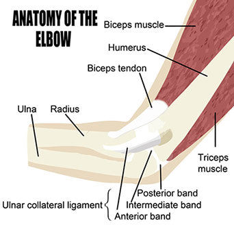
 Our elbow is a joint which is sometimes referred to as the funny bone. It connects the upper portion of the arm to the lower portion, or forearm. It is a synovial hinge joint due the unique arrangement of the bones which articulate to allow for some rotation as in addition to extension and flexion. These bones of the elbow are the humerus, ulna and radius.
Ligaments contribute to the stability of the elbow. Included in these are the medial collateral ligament, the lateral collateral ligament and the annular ligament and we will go into more details about each one shortly.
Important tendons attaching the muscle to bone in the elbow are the biceps and triceps tendons.
Muscles also work to form and operate the elbow. Muscles in the anatomy of the elbow are the brachialis, brachioradialis, biceps brachii, triceps brachii and pronator teres.
In order to gain a better understanding of the anatomy of the elbow and how these parts work in conjunction we’ll now take a more detailed look.
Bones involved in the Elbow Joint
The humerus of the upper arm meets with the ulna and radius of the forearm or lower arm in distinct articulations creating the connections or joints which we commonly refer to as the elbow.
Humuroulnar Joint is the connection of the trochlear notch of the ulna bone to the trochlear of the humerus bone. This is a hinge connection only allowing for simple extension and flexion.
Proximal radiolulnar Joint is where the head of the radius connects with the ulna. This connection allows for pronation and supination of the hand, ie. Face up or face down, during either flexion or extension.

Ligaments of the elbow
The ligaments of the below work to stabilize and support its mobility. Below you will find information about all of the major ligaments that effect the elbow.
- Medial Collateral Ligament sometimes called the Ulnar Collateral Ligament consists of two triangular bands, front and back.
- Lateral Collateral Ligament sometimes called the Radial Collateral Ligament is a short narrow band of ligament.
- Annular Ligament is a band of fibers that circle the head of the radius thereby keeping contact between the radius and the humerus.
See the collection of our Elbow braces.
Tendons of the Elbow
Biceps and triceps tendons are the two main tendons involved in the anatomy of the elbow. Tendons serve to attach muscle to bone.
- Biceps tendon attaches the biceps muscle to the radius bone. It lets the elbow bend with force.
- Triceps tendon attaches the triceps muscle on the back of the arm with the ulna. It lets the elbow straighten out with force (as in a push-up) This is a common culprit in the condition known as tennis elbow.
Muscles of the Elbow
The muscles of the elbow provide for its movement.
- Brachialis is the strongest elbow flexor when the palm of the hand is pronated (face up)
- Brachioradialis muscle flexes the elbow and aids in pronation (face up position) and supination (face down position).
- Biceps Brachii muscle’s role is to flex the elbow and and supinate the forearm.
- Tricepts Brachii is the main extensor of the elbow.
- Pronator Teres muscle aids in the flexion of the elbow and pronation of the forearm. These are the muscles involved in the injury known as golfer’s elbow.
In conclusion we can see that once again there are numerous essential components including bones, ligaments, tendons and muscles required to work together to create the very critical joint allowing optimal use of our arms and hands which we call our elbow.
Related Article: 3 Best Elbow Braces For Annoying Elbow Pain in 2020 (With Real Reviews)
Images (c) CanStockPhoto


Leave a comment